by Jesse Taub, Historian
The First Nuclear Magnetic Resonance Image (MRI)
MRI today is an essential technique for diagnosing a host of medical problems. It is based on the use of nuclear magnetic resonance (NMR) to analyze chemical substances. While many people contributed to making MRI the multi-billion dollar industry it is today, an advance that was made on Long Island had much to do with it. It was the first two-dimensional (2D) magnetic resonance image. Our Section’s History Committee is preparing an IEEE Historical Milestone proposal to honor this brilliant accomplishment. It will be sent to the IEEE History Committee for their approval. If our proposal is approved, it will be the third for Long Island. Previous ones were the Grumman Lunar Module and the First Blind Flight by Jimmy Doolittle at Mitchell field in 1929.
This first 2D image was accomplished by Paul Lauterbur, a Professor of Physical Chemistry at the University of Stony Brook. He documented his ideas in a notebook in 1971 and published results in Nature in 1973. Lauterbur was motivated to produce this image by his awareness of Dr. Raymond Damadian’s work at Down State Medical Center in Brooklyn. Damadian had observed that cancerous cells could be distinguished from normal ones by nuclear magnetic resonance (NMR) techniques. However, the only way to scan these cells was point by point. Lauterbur felt that generating a 2D image would make it practical for medical use.
Lauterbur realized that standard NMR measurements made with a fixed magnetic field, could not give precise distanced location of resonance. He reasoned that if he could linearly vary the magnetic field as a function of the distance, he could encode the data in one dimension. By rotating the varying field by three 45 degree steps, he was able to get enough data to describe a 2D image. He further realized that algorithms that had been used to generate X-ray CT scans could be applied to his problem. Furthermore, he reasoned that a series of 2D images (slices) could yield 3Dimage information.
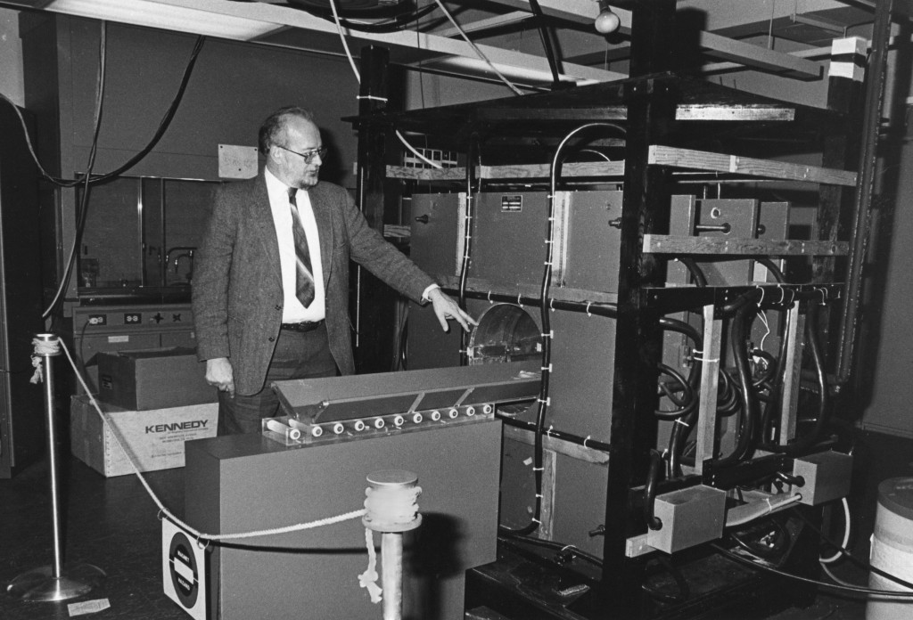
His results stimulated Peter Mansfield of the University of Nottingham, in the UK, to make improvements in the image quality and signal processing speed. Both of them received the Nobel Prize for Physiology or Medicine in 2003 for their MRI imaging accomplishments. Lauterbur’s original equipment, a Varian A60 NMR spectrometer that he added magnetic field gradients to, is housed in the lobby of the University of Stony Brook’s Chemistry building and is well worth looking at if you are in the area.